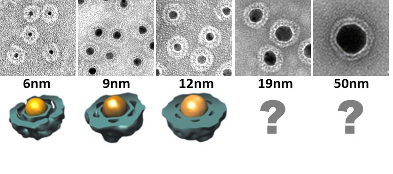
 Photothermal imaging of single nanoparticles is able to detect metal nanoparticles as small as 2.5 nm and to overcome the challenges associated with photophysical phenomena limiting the temporal dynamic range of other imaging methods. While in recent years fundamentals of image formation and instrumentation development have been relatively well covered thus offering a robust platform from the perspective of experimental control, the potential of photothermal microscopy in chemical imaging has remained relatively little explored.
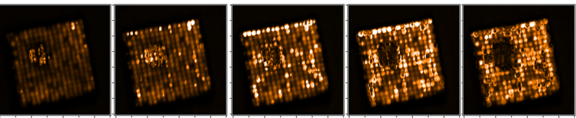
Our projects seek an improved understanding and control of phenomena involved in single nanoparticle photothermal imaging. We plan to apply this understanding to further our knowledge in the mechanisms of nanoparticle-initiated photochemistry. Specific projects starting this year include dynamics of heterogeneously nucleated phase transitions far from equilibrium, and in singulo growth kinetics of seeded bimetallic nanoparticles.
Remarkably, although assembled from hundreds to thousands of proteins, the capsids of many viruses from all domains of life are often perfectly monodisperse. Such capsid architectures are symmetric, vivid examples of spherical crystallography. The mechanisms by which such large, non-covalent systems achieve precise stoichiometry remain obscure. By analogy with the 3D crystal state, they raise questions fundamental to materials science: Do virus shells have different phases? What type of defects are encountered in lattices occupying a spherical manifold and what is their role in collective phenomena such as virus deformation or disassembly? How do virus shells grow to such precise stoichiometry? Focusing on the physical basis of function, these questions are important for establishing a solid foundation for design principles of virus-inspired materials.
![]() Copyright © 2020 | The Trustees of Indiana University | Last updated December 9, 2020 | Webmaster
Copyright © 2020 | The Trustees of Indiana University | Last updated December 9, 2020 | Webmaster